
Educational Models
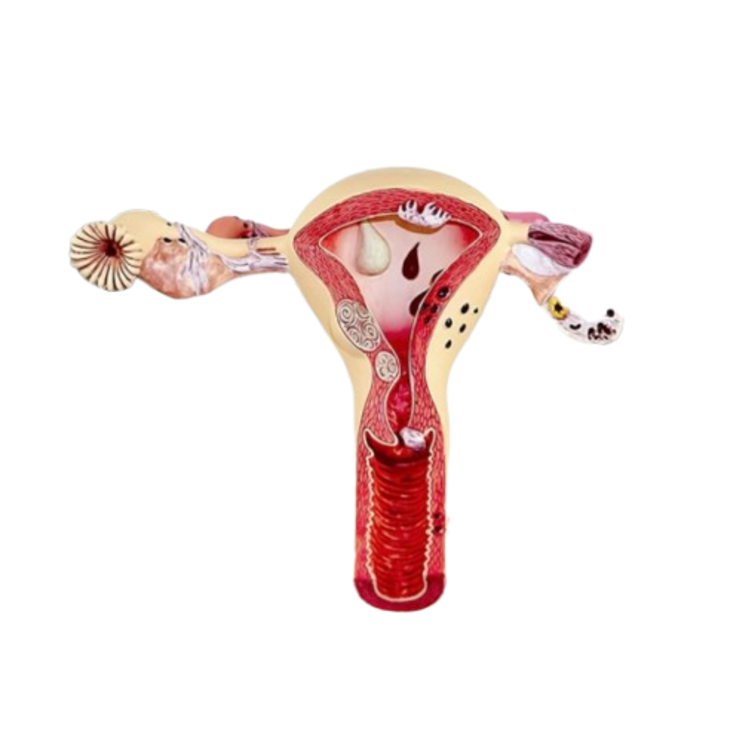
Models with uterus, fallopian tubes, ovaries
Specification:-
Models with uterus, fallopian tubes, ovaries
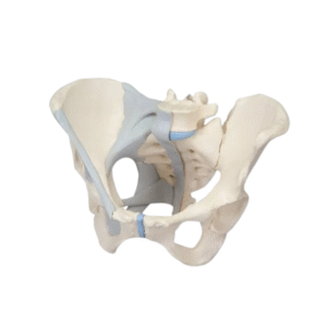
• Female pelvis with ligaments, vessels, nerves, pelvic floor and organs, in six pieces and offering detailed information on the topography of the bones,
ligaments, vessels, nerves, pelvic floor muscles and pelvic organs.
• Model showing the entire pelvic floor with the external anal sphincter, external urethral sphincter, the deep and superficial transverse perineum and
the partially removable bulbospongiosus and sectioned at the mid-sagittal level.
• The rectum, the uterus with the fallopian tubes, the ovaries and vagina must be removable and disassembled into two halves per section mid-sagittal
• The right half of the pelvis should show the divisions and topographic anatomy of the artery common iliac, external and internal artery, as well than
the common iliac vein and the vein external iliac. The right sacral plexus, the sciatic nerve right and the right internal pudendal nerve must also be
shown.
• Bones and ligaments presented: two iliac bones, the pubic symphysis, sacrum and coccyx, as well as the fifth lumbar vertebra with disc
intervertebral. A mid-sagittal section, passing by the 5th lumbar vertebra, the sacrum and the coccyx must allow the two halves of the pelvis,
revealing part of the ponytail in the vertebral canal. The left half of the 5th lumbar vertebral body is removable. Half right of the model should show
pelvic ligaments
following:
• inguinal ligament, sacrotuberous ligament, sacrospinal ligament, sacroiliac ligaments anterior, iliolumbar ligament, ligament anterior longitudinal,
sacroiliac ligament interosseous, posterior sacroiliac ligament and sealing membrane.

Reviews
There are no reviews yet.